|
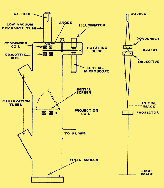
Explanatory sketch showing an electron microscope in simplified form. On the right is an electron-optical diagram to illustrate its operation.
Now that television is a going concern one is beginning to get used to optical terms encroaching on our preserve. A few years ago the idea of focusing by adjusting potentiometers would have seemed very odd both to the photographer and the radio-man. But when one actually sees the same result on a screen as the photographer does when he moves his lenses to and fro, there is obviously some close connection between the two. That being so, there should now be nothing very strange in the idea of an electrical microscope.
The ordinary optical microscope has gone about as far as it can, and that not very far. Sam Weller had doubts even about his pair o patent double million magnifyin gas microscopes of hextra power. The trouble is not so much that it is impossible to magnify things more, but that nothing is gained by doing so. It is the same sort of thing as with television pictures. Even if the picture could be made as big as a house it wouldnt reveal any more detail, because the ability to show detail - technically known as the resolving power - is limited by the number of lines in the television system. The resolving power of a microscope is limited by the wavelength of the light used. Although the wavelength of visible light is very small compared with even the shortest radio waves, it is no smaller than some of the tiny things one wants to see, and it is impossible to see clearly a detail that is much smaller than the wavelength of the light. So the useful magnification with visible light is limited to about 500. That is really more than it may seem because it is magnification of length, the corresponding magnification of the area of an object is, of course, 250,000.
You may have noticed that I said visible light. Some time ago it would have sounded like saying wet water or lifeless death, and, no doubt, would have provoked similar rude remarks about tautology or redundancy. But light is now generally understood to cover electromagnetic radiation not only within the band of wavelengths that is visible but also the invisible radiation of longer wavelength (infra-red) as far as the boundary of the band described as heat, and of shorter wavelength (ultra-violet) as far as the next known band - l forget what it is but it is a long way off. Obviously for extending the power of the microscope ultra-violet light offers possibilities, and is actually used to some extent, but the band that can be generated and detected in practice is quite narrow and only a slight advantage is gained.
There is a device called the ultra-microscope for detecting specks much smaller than the wavelength of light, in much the same way as specks of dust invisible to the naked eye can be seen in a beam of sun-light, but it is unable to disclose shape or form.
Greater Resolving Power
The development of cathode-ray tubes, particularly the specialised sorts used in television cameras, has shown that in many ways a ray of electrons can be manipulated like a beam of light, using either electrostatic deflecting plates or magnetic coils in place of lenses. The important difference is that the wavelength is thousands of times smaller and the theoretical resolving power therefore thousands of times greater, opening up new possibilities tor examining the minutenesses of nature.
The technique of generating and controlling rays of electrons and rendering them visible on a screen is all ready to hand, but there is one rather awkward difference between electrons and light. There is no difficulty in finding a substance glass through which light can easily pass when the thickness is amply great enough not only to bear the tiny weight of the objects to be examined but also to stand handling. But to allow an electron beam to pass, the slide must be of even less than gossamer thickness, otherwise it would not only stop the beam but be destroyed by it. Another thing that makes the electron microscope awkward for amateur microscopists is that the object has to be put inside the cathode ray tube, which means having handy all the apparatus for pumping the tube to a high vacuum. Still another limitation is that objects cannot be examined by the reflected beam as with light.
In spite of these drawbacks the electron microscope is very valuable for revealing particles that are quite invisible under the most powerful light microscope, and for showing the distinctive forms of microbes or powders that are otherwise only seen as undefinable specks. A magnification of 100,000 has already been claimed.
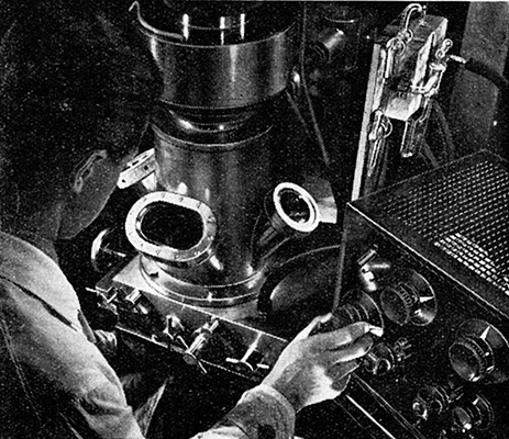
The photograph shows the Siemens electron microscope. It is a modified form of a television cathode-ray tube. A much higher anode voltage is used-something like 100,000 Volts. The beam is first made to converge on the object, which is supported on an incredibly thin film of collodion. Collodion is the stuff dissolved in ether that can be poured over a wound and is left as 'new skin' when the ether has evaporated. But this film has to be very much thinner, only about 0.00001 millimetre thick - and as it is naturally very delicate and fragile it is caught on a plate with a window that may be only a thirtieth of a millimetre in diameter. I should imagine that threading a No. 12 needle is as easy as hitting the side of a house compared with the job of getting the desired object exactly placed on this window. And if things are not properly arranged it is just too bad, because the vacuum has to be let down and re-pumped every time the slide is actually got at. Of course, if the object is on the collodion window at all the slide can be shifted by external adjustments to bring the desired part on to the screen. Even with a comparatively low-power microscope it may he quite difficult to find the place, and to make this easier in the electron microscope there is a screen for preliminary adjustment, at a position where the magnification is much less. After adjustment this screen is taken away and the final image appears on an enlarged scale.
The diagram shows in greatly simplified form a British electron microscope described by Martin, Whelpton and Parnum, and alongside is the electron-optical diagram. The electron beam is produced from a cold cathode in a low-vacuum tube at the top, and the hole in the anode is so small that it is possible to maintain a high vacuum in the main body of the apparatus. To help solve the problem of getting the desired object in the path of the beam it is placed on a revolving plate so that it can first be examined and adjusted by an optical microscope and then rotated into the electron beam. An image on a fluorescent screen (or photographic plate) is given at a moderate magnification half-way down for adjustment purposes; another stage of magnification yields the final image at the foot.
Apart from the obvious uses for the electron microscope, one wonders if it will be the means for extending our knowledge of the nature of matter and electricity. In a much simpler form it has been helpful in studying emitting surfaces - cathodes, in other words. In the early days valves depended on plain tungsten filaments run at bright white heat. Then came the thoriated dull-emitter filament. And now the oxide-coated cathode, run at a still lower temperature. The way in which emission takes place from these surfaces is of great interest to the valve and cathode-ray tube designer. By suitable adjustment of the electrode voltages it is possible to get an enlarged image of the cathode on a screen. If you have played about with the focusing of a torch or headlight you may have hit on an adjustment that projects an enlarged image of the lamp filament on the wall. An exactly analogous process gives this cathode image. I have watched a cathode being over-run and then rejuvenated, using this method. The bright parts of the picture are not necessarily the parts of the cathode that are hottest, they are those that are emitting most electrons. A spot of emissive coating, which might be seen as a dark patch on the cathode viewed direct, would appear on the screen as a patch of bright light on a dark ground.
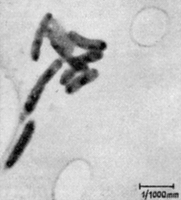
Illustration showing bacteria magnified 4, 200 times by the electron microscope and subsequently enlarged up to 10,000 times.
|