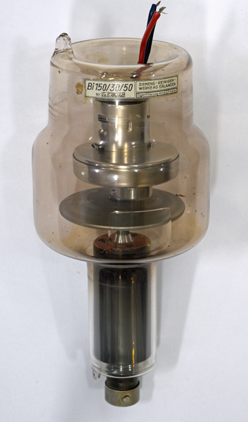
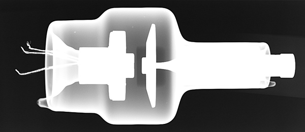
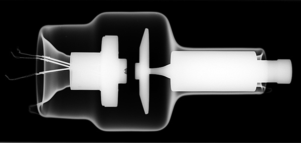
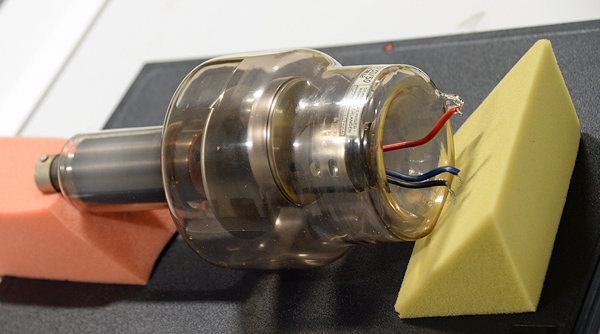
This exhibit is a rotating anode X-Ray tube with two filaments for fine and course focus. The lower part of the above image is the rotator that fits inside the coils placed around the neck of the tube. This motor spins the solid tungsten target so as to avoid local hot spots and allow for prolonged use.
See Also Mullard 1950's X-Ray tube in sectioned housing. for additional details.
The envelope glass has a window ground into it. This aims to provide a constant thickness of glass and be as thin as possible without risking the structural integrity of the tube.
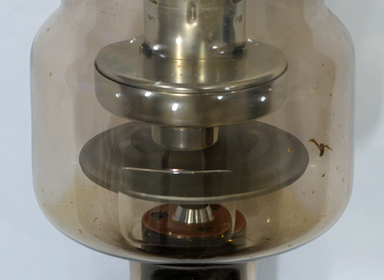
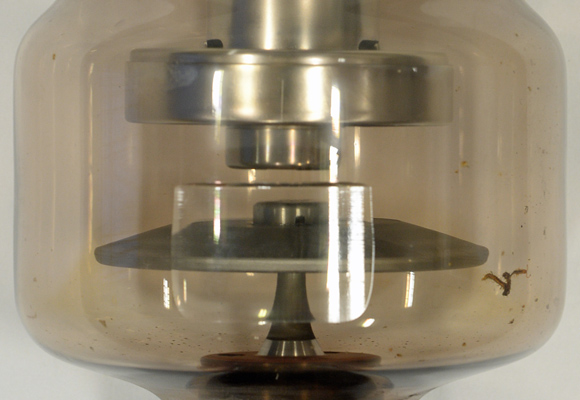
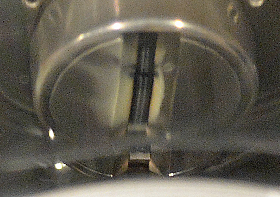
Looking through the exit window at the anode with the fixed filament cylinder above.
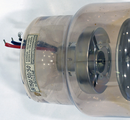
The filament end. The three wires allow for switching between filaments.
This X-Ray image shows the reentrant glass at both ends, thus placing strength close to the working elements. Note the darker rectangle of the exit window. It clearly provides some attenuation but less than the rest of the envelope.
Here the filaments are seen end on. The motor rotor comes close to the glass walls so as to maximise the effect of the rotating magnetic flux produced by the motor coils outside.
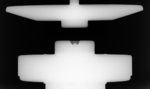
A closer view of the filaments.
Difficult to image properly due to the glass but here the two filaments can be seen.
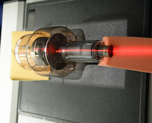
This image is of the tube on the bed of the X-Ray machine. The black rectangle is the image forming plate. This plate contains a chemical that forms a latent image when exposed to the X-Rays. Subsequent processing is by scanning with a red laser beam. This excitation 'develops' the image which appears in the ultra-violet. Filtration removes the red laser excitation and the UV is converted to an electrical signal by a photomultiplier. The resultant image is then available on a PC monitor or for export as a .JPG.
Alignment under the X-Ray head is by positioning to the red laser line.
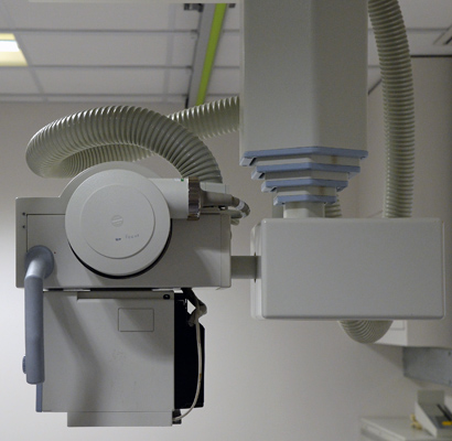
The X-Ray machine used to image the collection. The working source is contained within the cylinder. The high voltage lead from one end can be seen in the centre of the image.
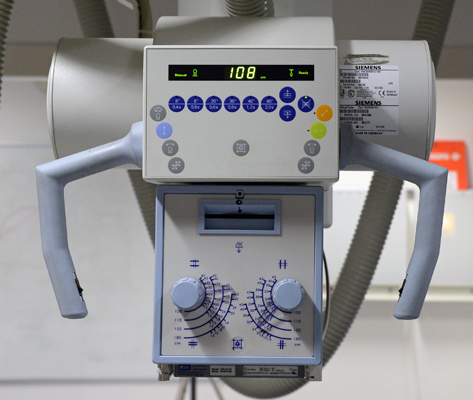
The control panel on the equipment. The label indicates that the X-Ray tube is Type Opti 150/30/50 HC.
The wide glass tube envelope is 140 mm in diameter, and including the base is 308 mm tall.